
Table of Contents
ToggleIntroduction to Microscopy
Microscopy is the scientific discipline of using microscopes to observe samples and items that are too small to be seen with the naked eye (objects that are not within the resolution range of the normal eye).
The light is typically concentrated on the sample by passing it through a condenser in order to achieve the highest possible intensity. The light passes through the sample, and then through the objective lens, which magnifies the image of the sample, before finally reaching the oculars, where the magnified image is seen.
Microscopic methods have advanced significantly over the last 20 years and are now an essential means of studying molecular processes at the subcellular level in order to obtain temporal and spatial data at high resolution. A solid grasp of cell biology, sample preparation, and fluorescence light microscopy are necessary for meticulously designing and executing microscopy-based experiments in order to get the best outcomes.
Types of Microscopes
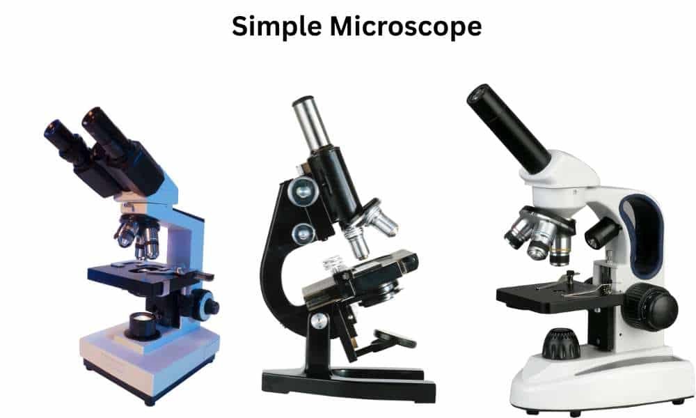
1. Simple Microscope
A simple microscope only has one lens, which magnifies objects. Due to the lens’s outward curve causing light rays to converge, the subject being seen seems larger and closer than it actually is. It usually magnifies between 2x and 20x, which is a little to medium amount. In 1670, Anton Van Leeuwenhoek created and conceived it.
Features
- Single Lens: A simple microscope only has one lens, which is typically convex in shape, such as biconvex or Plano convex.
- Magnification: These microscopes typically provide low to moderate magnification, ranging from 2x to 20x, depending on the lens being used.
- Cost: These microscopes are less expensive than others.
- Simple to Use: They are really simple to use. With this microscope, no specialized technical expertise is necessary.
Uses
- Applied to the study of insect, algal, and fungal morphology utilized to examine soil composition and type.
- Used for fixing watches, mobile phones, and other micro devices and components in electronic repair shops.
- Utilized by jewelers to assess the quality of rubies, diamonds, and other gemstones.
- Used for examining minutiae of engravings, manuscripts with smaller characters, and other things.
Limitations
- Have a really low magnification, up to 10 times.
- No mechanical stage and a mirror for illumination.
- Mandate that the stained sample be thin in order to provide a clear view.
- Extremely poor image resolution and contrast.
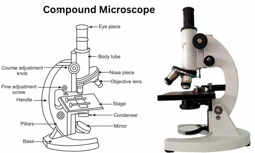
2. Compound Microscope
Compound The microscope is a kind of microscope that uses visible light for illumination and a system of several lenses to magnify the specimen. It usually has two lenses: an objective lens and an ocular lens. It can magnify pictures up to 100 times.
It is the most popular microscope used in biological disciplines such as medicine, microbiology, the life sciences, pathology, hematology, anatomy, molecular biology, and others.
The objective lens captures the light that passes through the specimen when it is placed on the stage and the light is concentrated through a condenser. At the body tube, an enlarged image is created. This is referred to as the main image. The ocular lens allows the light to pass through after it bends inside the body tube.
The image is magnified for the second time when it passes through the ocular lens. The secondary image is what this is known as. At last, a greatly magnified picture is produced at a distance of clear vision.
Types of Compound Microscopes
- Bright-field Microscope
- Dark-field Microscope
- Phase Contrast Microscope
- Fluorescence Microscope
Bright-field microscope: The Brightfield Microscope, sometimes referred to as the Compound Light Microscope, is an optical device that uses light rays to create a dark image against a bright background and is mostly used in biological research to examine stained samples.
Dark-field microscope: A darkfield microscope is a particular kind of optical microscope used in microscopy to see transparent specimens that would be challenging to observe using conventional bright-field illumination. The method works by shining light at an angle or slant onto the sample, which makes the specimen shine against a black backdrop. Particularly in samples that are almost transparent or have little inherent pigmentation, this method improves the contrast and visibility of minute features.
Phase Contrast Microscope: An optical microscope called a phase contrast microscope transforms minute phase variations in light into variations in light intensity, producing images with greater contrast that are easily seen by the human eye. A modest phase shift occurs when light passes through transparent materials, which is imperceptible to the naked eye. These minor phase variations are transformed into changes in the amplitude of light using phase plates. The difference in amplitude may be seen in the image’s contrast variations.
It can be used to see live cells in their native environment without fixing or staining them. With improved contrast, transparent specimens and subcellular organelles may be seen clearly.
When light passes through a sample, a slight phase shift occurs in the light rays because the specimen’s refractive index and thickness vary from one location to another. Variations in light intensity (brightness), which will increase the image’s contrast, can be converted from this phase change.
Fluorescence Microscopy: Light microscopy employing fluorescence is known as fluorescence microscopy. A substance is considered fluorescent if it absorbs the energy from invisible shorter wavelength radiation (like ultraviolet light) and releases longer wavelength radiation of visible light (like red or green light). Microorganisms, antibodies, and a wide variety of other compounds can be quickly identified using fluorescence, as it’s known, in clinical and diagnostic environments.
Because they contain fluorescent compounds like chlorophyll, certain cells naturally fluoresce under ultraviolet radiation. Fluorochromes are fluorescent colors that may be used to tint specimens that do not fluoresce naturally. Popular fluorescent dyes include; acridine orange, auraminerhodamine, DAPI (49, 6-diamidino-2-phenylindole), Alexa Fluors, or DyLight 488.
Uses
- Used in microbiology to investigate the shape of microbes.
- Used in histopathology to analyze tissues, cytopathic consequences, tumors, and other things.
- Used in cytology to examine the cellular architecture of various cell types.
- Used by scientists to view slides of cells, tissues, or portions of biological components.
Limitations
- Unable to image items smaller than the wavelength of visible light (0.4μm).
- Has a lower resolution and picture contrast.
- Not capable of seeing live internal structures.
- Need a sample that is thin and colored.
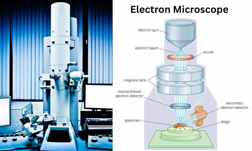
3. Electron Microscope
The atomic and molecular makeup and properties of materials may be effectively studied using electron microscopy. This involves using a focused stream of electrons to produce a high-resolution image of the specimen being examined.
This approach has transformed the way we see the world at its core and contributed significantly to progress in a wide range of fields, including biology and materials science.
Types of Electron Microscopes
- Transmission Electron Microscope (TEM)
- Scanning Electron Microscope (SEM)
- Environmental SEM (ESEM)
- Scanning Transmission Electron Microscope (STEM)
- Cryo-Electron Microscopy (Cryo-EM)
Microscopy Using Transmission Electrons (TEM): Sending an electron beam through a thin sample is the basis of the most widely used form of electron microscopy, TEM. The sample’s atoms and electron interactions produce a contrast image that can be used to visualize the sample’s internal structure. TEM has an amazing resolution and can produce images of structures as small as single atoms. It is commonly used in fields such as materials science, nanotechnology, and biology.
Scanning Electron Microscopy (SEM): Using a scanning electron microscope, or SEM, a concentrated beam of electrons is scanned over the surface of a sample. By utilizing detectors to collect the signals created when electrons interact with the atoms in the sample, an image of the sample’s surface may be produced. SEM is particularly helpful for photographing larger samples and can provide 3D information about the sample’s structure. It is frequently used in materials research, forensic investigation, and environmental sciences.
Environmental Scanning Electron Microscopy (ESEM): Materials can be imaged using ESEM in their untreated or hydrated states without needing any sample preparation. By maintaining a high-pressure environment inside the microscope, it allows for the imaging of specimens with high water content. ESEM is frequently used in the domains of biology, geology, and environmental science.
Scanning Transmission Electron Microscopy (STEM): The principles of TEM and SEM are combined in STEM, which scans a thin sample using a focused electron beam. Detectors can collect the signals produced as the electrons interact with the atoms in the specimen, and these signals can be used to create a picture of the specimen’s internal structure. STEM is commonly used in the fields of biology and materials research, and it may allow for high-resolution imaging of thin samples.
Cryo-Electron Microscopy (CryoEM): With cryo-EM, which doesn’t require staining or fixation, materials can be imaged in their hydrated, unaltered state. CryoEM technology allows for high-resolution imaging of large and complex structures, such as proteins and viruses, by rapidly freezing samples with liquid nitrogen while preserving their original structure. It is commonly employed in drug development and structural biology.
Uses
- Cutting-edge study in cell and molecular biology.
- The study of viruses and bacteria, among other microbes.
- Treating ailments effectively.
- High resolution imaging.
- 2d and 3d imaging.
- Pathology and the study of human anatomy.
- Disease diagnosis and treatment.
- Increased knowledge of germs and pathogens.
Limitations
- It is extremely sensitive equipment that is costly and takes up a lot of room in the lab.
- In order to prevent arti-facts and ensure correct use of the microscope, trained labor is required for proper sample preparation.
- Live specimens cannot be viewed since electron microscopes function in a vacuum.
- Because electron beams have poor penetration capabilities, the subject needs to be very thin. Before observation (for TEM), the sample is dried and sliced into ultrathin pieces. This may result in structural distortions.
- Their functioning may be impacted by vibrations and magnetic fields produced by other laboratory apparatus.
4. Confocal Microscope
A confocal microscope employs a spatial pinhole to block out-of-focus light and only uses light from the plane of focus to create a 3D image with improved resolution and image contrast. Additionally, it goes by the name confocal laser scanning microscope.
A fluorescence microscope of this kind is used to create two- or three-dimensional images of samples that are comparatively thick. With this method, the excitation light is concentrated on a particular area of the specimen that is on the focal plane. The focal point is optically altered to scan the whole sample and produce a three-dimensional picture. A high-resolution image with improved contrast is produced.
Laser light is used for illumination in this kind of microscope. It was possible to concentrate the laser beam on a certain location thanks to the use of a confocal aperture and oscillating mirror. They block rapid photo bleaching and light scattering while ignoring background noise from unfocused regions.
The optical sectioning method, which combines several 2D images to create a 3D image, is the foundation of it.
Laser light is used to illuminate the fluorophore-stained specimen. The laser beam is directed onto the fluorophore-stained sample. The optical route includes a pinhole through which the emitted fluorescent light is transmitted. The focused point’s released light is allowed to pass through selectively, while all other background lights are blocked. A photomultiplier tube transforms light into an electrical signal. The electrical signal is analyzed by computer software, which generates a three-dimensional picture.
Working Principle of Confocal Microscope
- Laser excites fluorophores in the sample,
- A spatial pinhole blocks out-of-focus light,
- A photomultiplier tube converts emitted light into an electrical signal,
- Image is reconstructed using computer software,
Confocal Microscope
- Used to identify fungal cells in corneal scrapings and eye corneal diseases.
- Used for ensuring the quality of pharmaceutical goods.
- Used for optical 3D imaging and scanning.
Limitations
- High cost and complex setup.
- Limited to specific excitation wavelengths.
- Risk of photobleaching with prolonged exposure.
Applications of Microscopy in Microbiology
Microscopy is the foundation of microbiological research and diagnostics. Here’s how it’s used:
- Clinical Diagnosis: Identifying bacteria, viruses, fungi, and parasites in patient samples.
- Cell Biology: Observing cellular organelles, mitosis, and gene expression.
- Immunofluorescence: Detecting specific proteins or antigens using antibody staining.
- Virology: Visualizing virus particles with electron microscopy.
- Mycology and Parasitology: Examining fungal structures and parasitic forms.
- Industrial Microbiology: Monitoring fermentation, quality control in pharmaceuticals.
- Environmental Microbiology: Analyzing water, soil, and air microorganisms.
Comparison Table of Microscope Types
| Microscope Type | Light Source | Max. Magnification | Best For | Cost | Resolution |
| Simple | Natural/Artificial | 20x | Basic observation | Low | Low |
| Compound | Visible light | ~1000x | Biological studies | Moderate | ~0.2 µm |
| Fluorescence | UV/LED | ~1500x | Cell labeling | High | Moderate |
| Electron (TEM/SEM) | Electron beam | >1,000,000x | Ultrastructure | Very High | ~0.1 nm |
| Confocal | Laser | ~2000x (3D) | Fluorescent imaging | High | High |
Conclusion
Microscopy is one of the most essential tools in microbiology and modern science. From the simplicity of a single lens to the sophistication of electron and confocal systems, microscopes allow us to explore the microscopic world in unparalleled detail. These technologies support ground breaking research in disease mechanisms, molecular biology, materials engineering, and drug development.
Whether you’re a student, researcher, or healthcare professional, understanding the types of microscopes and their applications will help you unlock the power of the invisible world.
Also Read:
Reference and Sources
