Table of Contents
ToggleIntroduction to Staining in Microbiology
The act of staining cells, tissues, or tiny pieces gives them color, which improves their visibility and visualization under a microscope. Different staining techniques are employed for diverse applications, including the examination of metabolic processes, cytopathology, microscopic organisms, and cellular structures. Examples of such techniques are simple, differential, and special staining.
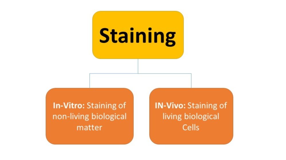
A substance called “stain” is used to perform the staining. The colourless specimen is coloured by this method, which makes use of a wide range of natural and synthetic stains.
The two methods for doing it are in vitro and in vivo, as described below:
Steps in the Staining Process
In general, the staining protocol comprises three consecutive steps:
- Smear preparation: This is the first step of the smear preparation process. It entails mixing the inoculum with a drop of sterile water and spreading it over the glass slide until a thin film is produced.
- Smear fixation: This is the second step, during which the thin layer of microorganisms that has formed on the glass slide is dried and heat-fixed.
- Specimen staining: In this last step, the stain is applied to the dried smear, giving the microscopic material its colour. Before biochemical experiments and microscopic analysis, this technique is performed.
By highlighting contrast between different components, stains allow detailed visualization of structures like nuclei, flagella, capsules, and spores.
Types of Stains Based on Chemical Nature
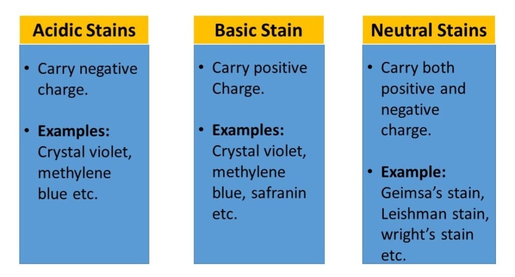
Stains are categorized into three types based on their chemical composition:
Acidic Stains:
- Carry a negative charge.
- Stain positively charged components like proteins.
- Example: Eosin.
Basic Stains:
- Carry a positive charge.
- Bind to negatively charged cell components like nucleic acids and bacterial cell walls.
- Examples: Crystal violet, Methylene blue, Carbol fuchsin.
Neutral Stains:
- Contain both acidic and basic dyes.
- Useful for staining complex cell structures like blood smears.
Categories of Staining Techniques
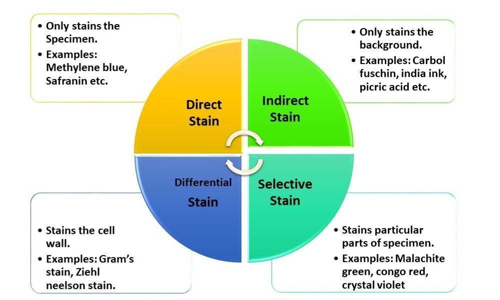
Based on purpose and application, staining techniques are classified into:
- Direct Staining – Stain directly applied to the microorganism.
- Indirect Staining – Background is stained, leaving organisms clear (negative staining).
- Differential Staining – Differentiates between microbial groups (e.g., Gram staining).
- Selective Staining – Targets specific structures like capsules, spores, or flagella.
Importance of Staining in Microbiology
- Staining aids in our ability to see the organism better: Microbes are extremely tiny organisms that are also transparent, thus staining makes the specimen easier to identify.
- Aids in distinguishing between organisms: Depending on the color retention capabilities of the cells, staining helps differentiate between the two distinct classes of organisms (some microbes retain the color of stain, while others don’t).
- To single out a specific structure: In order to learn more about microorganisms, it is also necessary to examine the numerous internal and external structures of organisms, such as flagella, capsules, nuclei, spores, etc.
Major Types of Staining Techniques
1. Simple Staining
Definition: Uses a single basic dye to color microorganisms.
Purpose: To determine the size, shape, and arrangement of bacterial cells.
Common dyes used:
- Methylene blue
- Crystal violet
- Carbol fuchsin
Procedure:
- Prepare and fix the smear.
- Flood the slide with dye for 30–60 seconds.
- Wash with water and blot dry.
- Observe under microscope.
2. Differential Staining
The purpose of these staining methods is to differentiate between organisms according to their staining characteristics.
For example: Gram staining, which separates bacteria into two groups—Gram negative and Gram positive—is a bit more complex than basic staining procedures, which may expose the cells to several dyes or stains.
There are many different varieties of differential staining, including Gram staining and Acid Fast Staining.
A. Gram Staining
The Gram stain, developed by Christian Gram (1884), is the most widely used technique.
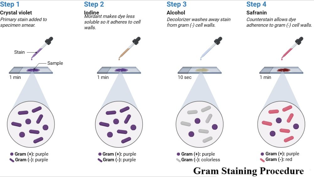
- Gram-positive bacteria: Thick peptidoglycan wall, retain crystal violet-iodine complex → appear purple.
- Gram-negative bacteria: Thin peptidoglycan, high lipid content, lose primary stain during decolorization → appear pink/red after counterstain.
Procedure:
- Make a bacterial smear on a clean glass slide, let it air dry, and heat fix it by running it through a flame.
- Give the slide a crystal violet stain, let it sit for one minute, and then rinse it with water.
- To fix the initial stain, add Gram’s iodine solution for a minute, then rinse with water.
- Use 95% alcohol or acetone alcohol to decolorize for 10 to 20 seconds, and then rinse right away with water.
- As a counterstain, apply safranin stain for 30–60 seconds before rinsing with water.
- Using absorbent paper, dry the slide.
- Use oil immersion and a microscope to view at a magnification of 100x.
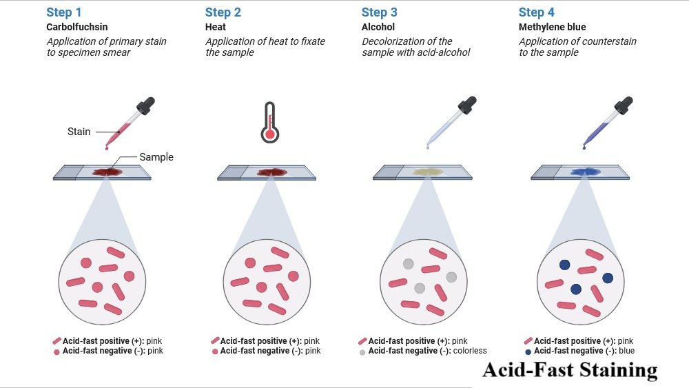
B. Acid-Fast Staining
Acid fast Staining is used for organisms like Mycobacterium tuberculosis, which have waxy mycolic acids in their cell walls.
Steps:
- Apply carbol fuchsin with heat.
- Decolorize with acid-alcohol.
- Counterstain with methylene blue.
- Acid-fast organisms → red
- Non-acid-fast organisms → blue
3. Special Staining
Certain staining methods employ particular dyes or methods to emphasize certain cell structures, compounds, or microorganisms. These methods are employed in in-depth research of specific components of the cell.
The following are a few of these staining techniques:
(a) Capsule Staining
- Capsules are non-ionic polysaccharides, resistant to staining.
- A combination of acidic (background) and basic (cell) stains are used.
- Capsule remains colorless, visible as a halo.
(b) Endospore Staining
- Endospores are highly resistant dormant structures.
- Stained using malachite green with heat, then counterstained with safranin.
- Spores → green, Vegetative cells → red
(c) Flagella Staining
- Flagella are extremely thin and delicate.
- Special mordants plus stains like carbol fuchsin make them visible.
- Useful for studying motility and bacterial classification.
Applications of Staining Techniques
- Medical microbiology: Diagnosis of infectious diseases.
- Environmental microbiology: Identifying microbes in soil and water.
- Food microbiology: Detecting contamination.
- Research: Studying morphology, physiology, and microbial structures.
Conclusion
In conclusion, staining procedures are crucial to microscopy because they allow us to see and identify microorganisms, cellular structures, and other minute samples. Numerous staining techniques, such as simple, differential, and special staining, offer important insights into the morphology, makeup, and features of cells and microorganisms. Researchers may learn about the makeup, purpose, and conduct of microbes by using various stains and methodologies, which will eventually aid in progress in fields such as medicine, biology, and microbiology.
FAQs on Staining Techniques
Q1. Why is staining important in microbiology?
Staining provides contrast, making microorganisms visible and identifiable under the microscope.
Q2. What is the difference between simple and differential staining?
Simple staining uses one dye to highlight morphology.
Differential staining uses multiple dyes to distinguish groups (e.g., Gram-positive vs. Gram-negative).
Q3. Which organisms require acid-fast staining?
Mycobacterium species, such as M. tuberculosis and M. leprae, which have waxy lipid-rich cell walls.
Q4. What is the purpose of capsule staining?
To visualize bacterial capsules, which act as protective virulence factors.
Q5. Can live cells be stained?
Yes, in vivo stains can be used to observe live cells without killing them, although most microbiological stains are applied after fixation.
Q6. What dye is used for endospore staining?
Malachite green is the primary stain, with safranin as the counterstain.
Also Read
- HIV and AIDS: Structure, Transmission, Symptoms, Diagnosis and Prevention
- Single Cell Protein (SCP): Sources, Production, Advantages, Disadvantages & Applications
- Detection of Viruses: Modern Methods, Applications, and Advancements
- Molecular Blotting: Techniques, Uses, and Significance
- Mycorrhiza: Structure, Types, and Role in Plant Growth
- Spirulina: The Superfood Microalga with Limitless Potential
- Immunology Quiz
- Microbiology Notes
- Bacteriology Quiz

